
Fig. 5
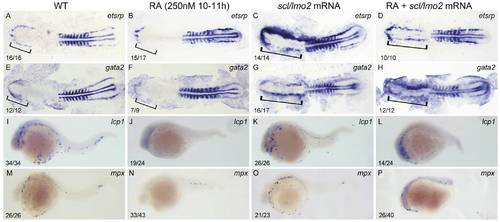
Overexpressions of scl and lmo2 into RA-treated zebrafish embryos partially rescue the defective primitive myelopoiesis. Both flat-mounted embryos (A–H) and whole-mounted embryos (I–P) are positioned anterior left and dorsal front (A–H) or lateral front (I–P). Embryos were treated with vehicle DMSO (A, E, I, M), 250 nM RA during 10 to 11 hpf (B, F, J, N), or microinjected with scl and lmo2 mRNA at 1–2-cell stage (C, G, K, O), or microinjected with scl and lmo2 mRNA at 1–2-cell stage and then treated with 250 nM RA during 10 to 11 hpf (D, H, L, P), respectively. They were then examined for expressions of hemangioblast markers etsrp (A–D) and gata2 (E–H) at 14 hpf, and myeloid markers lcp1 (I–L) and mpx (M–P) at 24 hpf by whole mount in situ hybridization. Expression of myoD in somites was used for staging (A–H). Bracket indicates the location of RBI (A, B, E, F), and ALPM (C, D, G, H). The number shown in the lower left-hand corner of each panel is the number of embryos exhibiting the typical phenotype shown in the panel to the number of embryos totally observed. |