
Fig. 4
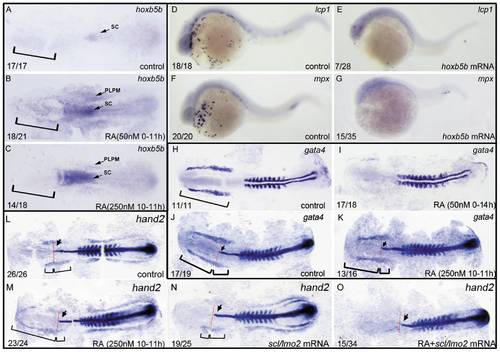
ALPM is lost in the embryos treated with 50 nM RA from 1–2-cell stage to 11 hpf but not eliminated in the ones treated with 250 nM RA during 10–11 hpf. All embryos including flat-mounted embryos (A–C, H–O) and whole-mounted embryos (D–G) are positioned anterior left and dorsal front (A–C, H–O) or lateral front (D–G). Embryos treated with vehicle DMSO (A), 50 nM RA from 1–2-cell stage to 11 hpf (B), and 250 nM RA during 10–11 hpf (C) were examined for hoxb5b expression at 11 hpf (A–C). Embryos treated with 50 nM RA treatment from 1–2-cell stage to 11 hpf displayed ectopically expression of hoxb5b in ALPM (B) but the ones treated with 250 nM RA during 10–11 hpf did not exhibit this ectopical expression (C). Compared with control embryos (D, F), overexpressions of hoxb5b by microinjecting embryos at 1–2-cell stage with hoxb5b mRNA significantly suppressed expressions of lcp1 (E) and mpx (G) at 24 hpf. Embryos treated with vehicle DMSO (H, J), 50 nM RA from 1–2-cell stage to 14 hpf (I) or 250 nM RA during 10–11 hpf (K) were examined for expressions of ALPM marker gata4 at 14 hpf. The location of ALPM at 11 hpf (A–C) or at 14 hpf (H, J, K) is indicated by bracket. Embryos treated with vehicle DMSO (L), 250 nM RA during 10–11 hpf (M, O), or microinjected with scl/lmo2 mRNA (N, O) were examined for expressions of cardiac marker hand2 at 14 hpf. Expression of myoD in somites (H–O) was used for staging and ntl expression was used for labeling embryonic axial mesoderm (J–O). The length between the anterior end of gata4 expression domain (J, K) or hand 2 expression domain (L–N) and the anterior end of ntl expression domain and the length between the posterior end of gata4 expression domain (J, K) or hand 2 expression domain (L–N) and the anterior end of ntl expression domain are marked by two different brackets. Red line denotes the anterior level of ntl expression domain (J–O). Arrow indicates the most anterior end of notochord marked by expression of ntl (J–O). sc: spinal cord; PLPM: posterior lateral plate mesoderm. The number shown in the lower left-hand corner of each panel is the number of embryos exhibiting the typical phenotype shown in the panel to the number of embryos totally observed. |