
Fig. S6
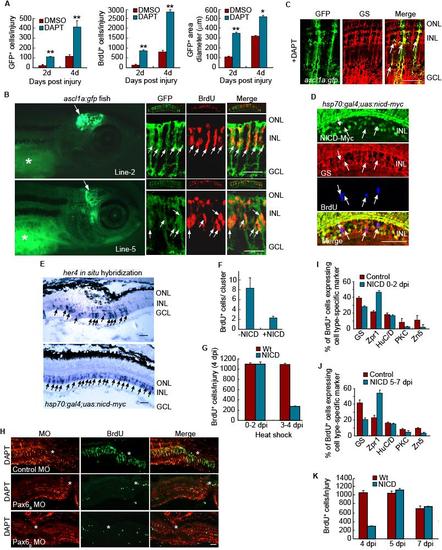
Notch and Pax6-dependent regulation of MG dedifferentiation and retina regeneration. Related to Figure 5. (A) Quantification of GFP+ and BrdU+ cells, along with the area they occupy shown in Figure 5A. **P<0.01, *P<0.05. (B) Four different lines of ascl1a:gfp transgenic fish were generated with each exhibiting transgene expression in the developing midbrain. (2 examples shown in left hand panels). * identifies autofluorescence in the yolk. Right-hand panels show sections through the adult injured retina where transgene expression is confined to BrdU+ progenitors. The small top inset for each panel shows a low magnification view of the retina. (C) In the adult injured retina of ascl1a:gfp fish treated with DAPT, transgene expression is confined to glutamine synthetase (GS)-expressing MG-derived progenitors. Scale bar, 50 μm. (D) NICD-myc, GS and BrdU immunofluoresence at 4 dpi shows NICD over-expression in the injured retina inhibits progenitor proliferation and the few remaining BrdU+ cells do not express NICD. (E) In situ hybridization assays show NICD over-expression stimulates her4 expression throughout the inner nuclear layer. (F) NICD overexpression reduces the number of BrdU+ cells in a neurogenic cluster. (G) NICD overexpression from 0-2 dpi has little effect on progenitor proliferation while NICD overexpression from 3-4 dpi suppresses progenitor proliferation. (H) Pax6a or Pax6b knockdown suppresses DAPT-dependent proliferation of MG-derived progenitors assayed at 4 dpi. (I) Retinal cell types regenerated at 14 dpi following NICD overexpression from 0-2 dpi. (J) Retinal cell types regenerated at 14 dpi following NICD overexpression from 5-7 dpi. (K) Proliferating progenitors remaining after NICD overexpression from 0-4 dpi can expand and contribute to retina regeneration at later times. Error bars are standard deviation. Scale bar, 50 μm. |
Reprinted from Developmental Cell, 22(2), Wan, J., Ramachandran, R., and Goldman, D., HB-EGF Is Necessary and Sufficient for Müller Glia Dedifferentiation and Retina Regeneration, 334-347, Copyright (2012) with permission from Elsevier. Full text @ Dev. Cell