
Fig. S4
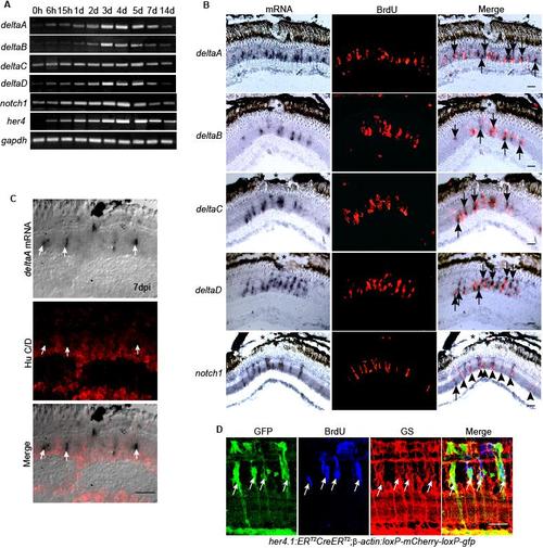
Notch signaling components are induced in proliferating MG-derived progenitors residing in the injured retina. Related to Figure 4. (A) RT-PCR showing the temporal pattern of Notch signaling component gene expression. (B) In situ hybridization combined with BrdU immunofluorescence shows that at 4 dpi genes encoding the Notch ligands, deltaA-D are restricted to the site of injury (*) and are predominantly localized adjacent to BrdU+ progenitors, while Notch receptor, notch1 is predominantly expressed in BrdU+ progenitors at the injury site. (C) In situ hybridization and HuC/D immunofluorescence show deltaA expressing cells are HuC/D-. (D) her4.1:ERT2CreERT2;β-actin:loxP-mCherry-loxP-gfp double transgenic fish show that the Notch responsive promoter, her4, is activated in proliferating MG-derived progenitors identified by BrdU incorporation and glutamine synthetase (GS) expression. Scale bar, 50 μm. |
Reprinted from Developmental Cell, 22(2), Wan, J., Ramachandran, R., and Goldman, D., HB-EGF Is Necessary and Sufficient for Müller Glia Dedifferentiation and Retina Regeneration, 334-347, Copyright (2012) with permission from Elsevier. Full text @ Dev. Cell