
Fig. 4
- ID
- ZDB-FIG-120216-15
- Publication
- Parker et al., 2011 - Ancient Pbx-Hox Signatures Define Hundreds of Vertebrate Developmental Enhancers
- Other Figures
- All Figure Page
- Back to All Figure Page
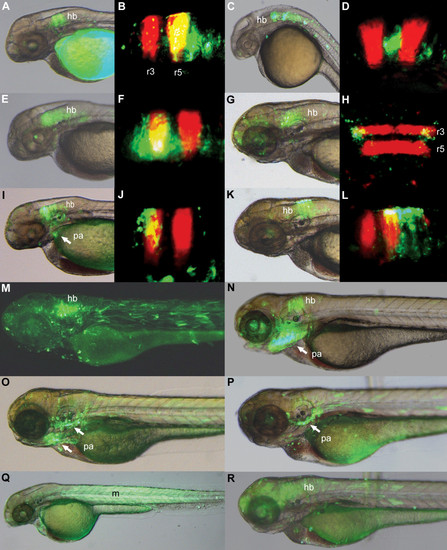
Pbx-Hox motifs correlate with segment-specific hindbrain and pharyngeal arch reporter expression. A-R, zebrafish elements from the lamprey (A-J, M, O, Q) and jawed vertebrate (K, L, N, P, R) CNE sets drive GFP expression in the hindbrain and pharyngeal arches. Elements: Evi1_40224 (A, B), Tshz3_43509 (C, D), NR2F2_27254 (E, F), Pax2_217 (G, dorsal view: H), ZNF503_32799 (I, J), Nkx6-1_4281 (K, L), Tshz3_24804 (M), Pax9_2099 (N), TshZ3_24805-6 (O), FoxP1_886 (P), Tshz3_24807 (Q), BCL11A_2554 (R). Expression in the hindbrain is often restricted to certain rhombomeres, as shown by comparison with r3r5 RFP expression (B, D, F, H, J, L). Tshz3_24807 drives expression in the trunk musculature (Q). Elements show temporal variation in reporter expression, expressing most strongly at 24-30hpf (C, D), 48-54hpf (A, B, E, F, I, J, Q) or 72-78hpf (G, H, K, L, M, N, O, P, R). hb: hindbrain; pa: pharyngeal arches; m: muscle. |