
Fig. 5
- ID
- ZDB-FIG-120214-3
- Publication
- Malicki et al., 1996 - Mutations affecting development of the zebrafish retina
- Other Figures
- All Figure Page
- Back to All Figure Page
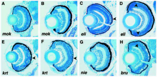
Phenotypes of mutations predominantly affecting development of the outer retina analyzed on histological sections (A) In mikre oko (mok)m632, a poorly differentiated photoreceptor cell layer is present in the central retina at 3 dpf. The presumptive photoreceptor cells frequently do not display the wild-type, elongated shape. (B) At 5 dpf, the photoreceptor cell layer is absent in mokm632. (C) The elipsa (eli)m649 retina at 3 dpf. Excessive cell death is most obvious in the central portion of the photoreceptor cell layer (arrowhead in C). (D) At 5 dpf most of the photoreceptor cells in elim649 are absent. Some cells survive in the periphery of the photoreceptor cell layer (arrowheads in D). (E) The krenty (krt)m699 retina at 3 dpf. The photoreceptor cell layer is discontinuous (arrowhead in E). Gaps in the array of photoreceptor cells appear to be filled with cells originating from the inner nuclear layer. This pattern persists till 5 dpf (F). (G) Retina of niezerka (nie)m743 at 5 dpf. The photoreceptor cell layer is missing. (H) brudas (bru)m148 retina at 3 dpf. As in elim649 some morphologically normal photoreceptors are present in the periphery (arrowhead in H). All sections are transverse. In all panels ventral is down. |
| Fish: | |
|---|---|
| Observed In: | |
| Stage Range: | Protruding-mouth to Day 5 |