
FIGURE
Fig. S2
Fig. S2
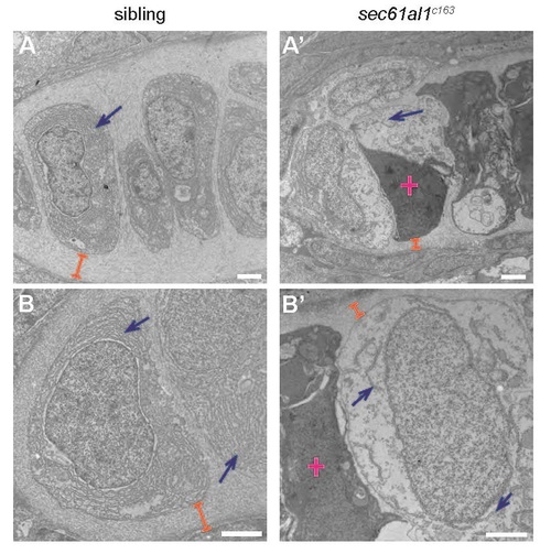
Transmission electron micrographs of jaw chondrocytes show ER and ECM deficits in sec61al1c163 mutants. (A, B) Chondrocytes of WT controls have dense ER throughout the cytoplasm (blue arrows) and extensive ECM surrounding chondrocytes (orange brackets). (A′, B′) Endoplasmic reticuli are sparse and discontinuous in sec61al1c163 mutants and the ECM is reduced in thickness. Dead cells are common in sec61al1c163 mutant micrographs (pink cross). All scale bars = 2 µm. |
Expression Data
Expression Detail
Antibody Labeling
Phenotype Data
| Fish: | |
|---|---|
| Observed In: | |
| Stage: | Protruding-mouth |
Phenotype Detail
Acknowledgments
This image is the copyrighted work of the attributed author or publisher, and
ZFIN has permission only to display this image to its users.
Additional permissions should be obtained from the applicable author or publisher of the image.
Reprinted from Developmental Biology, 360(1), Doll, C.A., Burkart, J.T., Hope, K.D., Halpern, M.E., and Gamse, J.T., Subnuclear development of the zebrafish habenular nuclei requires ER translocon function, 44-57, Copyright (2011) with permission from Elsevier. Full text @ Dev. Biol.