
Fig. 8
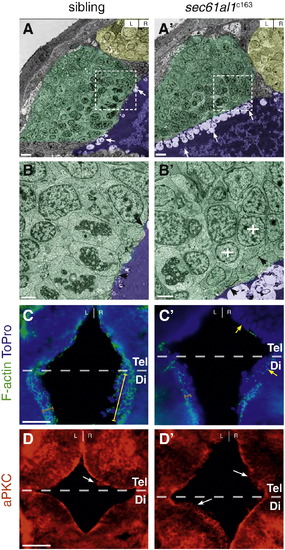
The ventricular epithelium is disrupted in sec61al1c163 mutants. (A) Transmission electron micrographs from coronal sections through the dorsal diencephalon of WT and (A′) sec61al1c163 mutant larvae at 54 hpf, with the pineal organ (yellow), habenulae (green), and 3rd ventricle (purple) indicated by false coloring. (B, B′) Higher magnification shows significantly more apical blebbing from cells lining the ventricle in mutants compared to their siblings (arrows). (B) The well-defined columnar morphology and cell–cell junctions (black arrowheads) of WT ventricular cells are not found (B′) in mutants. Rather, cells proximal to the ventricle are rounded (white crosses) and have few cell–cell junctions (black arrowheads). (C) Filamentous actin (F-actin) in the ventricular neuroepithelium localizes to the apical membrane (orange bracket) in cells along the entire rostro-caudal axis of the anterior 3rd ventricle. (C′) sec61al1c163 mutants have reduced (orange bracket) F-actin in the caudal ventricular neuroepithelium (orange bracket) and many cells without apically-localized F-actin in the rostral ventricular neuroepithelium (yellow arrowheads). (D) aPKC localizes to the apical surface of the ventricular neuroepithelium in wild type embryos, whereas (D′) apical aPKC is reduced or absent in many cells of mutants. C, D are from 10 μm thick cryosections, taken from the dorsal aspect. F-actin sections (C) are roughly 30 μm ventral from the dorsal surface, and aPKC sections (D) roughly 10 μm from the dorsal diencephalon. Tel, telencephalon; Di, diencephalon. Dashed boxes in A, A′ correspond to the region of higher magnification in the adjacent micrographs, B, B′. Scale bars: A,A′ = 20 7mu;m; B,B′ = 5 μm; C–D′ = 20 μm. |
| Fish: | |
|---|---|
| Observed In: | |
| Stage: | Long-pec |
Reprinted from Developmental Biology, 360(1), Doll, C.A., Burkart, J.T., Hope, K.D., Halpern, M.E., and Gamse, J.T., Subnuclear development of the zebrafish habenular nuclei requires ER translocon function, 44-57, Copyright (2011) with permission from Elsevier. Full text @ Dev. Biol.