
Fig. S1
- ID
- ZDB-FIG-120126-7
- Publication
- Peukert et al., 2011 - Lhx2 and lhx9 determine neuronal differentiation and compartition in the caudal forebrain by regulating wnt signaling
- Other Figures
- All Figure Page
- Back to All Figure Page
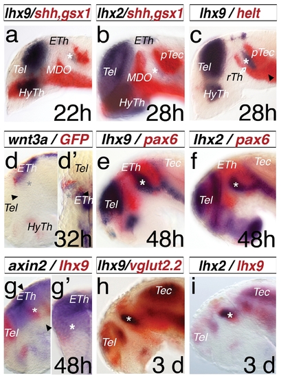
Expression pattern of lhx2 and lhx9 during thalamus development. A double in situ hybridization approach was used for analysis. All embryos were mounted laterally with stages indicated, except (d2) is a dorsal view and (g2) is a cross-section of the left hemisphere. lhx9 reveals an onset of expression in the thalamus (Th) at 22 hpf (a, asterisk), limited anteriorly by shh, a marker of the MDO and posteriorly by gsx1, a marker of the pretectum (PTec). At 28 hpf, lhx2 shows an onset of expression in the thalamus (b, asterisk). Within the thalamus, at 28 hpf helt marks the rostral thalamus (rTh) and the pretectum (c), however the lhx9 expression domain shows no overlap with the helt domain. The epithalamus is marked by the Wnt ligand, wnt3a, and the expression of the Wnt reporter 7×TCF-siam:GFP (d). The dorsal view reveals lateral a stronger expression of gfp-mRNA in comparison to the wnt3a pattern (d2). At 48 hpf, lhx2 and lhx9 show specific expression patterns in the telencephalon (Tel), thalamus (asterisk), and ventral to the tectum (Tec), indicated by the overlapping expression domain of pax6a, marking the alar plate of the forebrain during development (e, f). axin2 expression in the thalamus co-localizes with the lhx9 expression. (g, g2). vglut2.2, a marker of glutamatergic neurons in the relay thalamus (cTh), shows an overlapping expression domain with lhx9 (h). Both genes, lhx2 and lhx9, mark the thalamus at 3 dpf (i). ETh, epithalamus; HyTh, hypothalamus; MDO, mid-diencephalic-organizer; PTec, pretectum; RP, roof plate; rTh, rostral thalamus; Tec, tectum ; Tel, telencephalon. |