
Fig. 1
- ID
- ZDB-FIG-120126-1
- Publication
- Peukert et al., 2011 - Lhx2 and lhx9 determine neuronal differentiation and compartition in the caudal forebrain by regulating wnt signaling
- Other Figures
- All Figure Page
- Back to All Figure Page
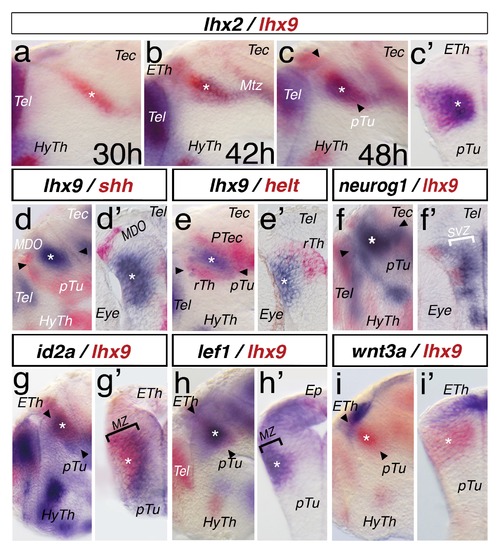
Dynamic expression pattern of lhx2 and lhx9 during regionalization of the caudal forebrain. A double in situ hybridization approach for thalamic development. Embryos were mounted laterally (a, b, c, etc.) or sectioned and the left hemisphere is shown (c2, d2, e2, etc.). Plane of section is indicated in the previous picture with black arrowheads. Asterisks mark the position of the thalamus. Marker genes and stages are indicated (a, b), all other embryos (c–i2) are 48 hpf. lhx2 expression is stained in red and lhx9 is stained in blue. lhx9 expression is revealed in the thalamus at 30 hpf (a). At 42 hpf, lhx9 expression increases and lhx2 expression is detectable ventro-posteriorly within the lhx9 domain (b). At 48 hpf, lhx2 and lhx9 overlap in the Th (c) and cross-section analysis reveals an overlap of both markers within the mantle zone of the thalamus (c2). The shh-positive mid-diencephalic organizer (MDO) is located anterior to the lhx9 positive thalamus (d), and a cryo-section reveals a gap between both expression domains (d2). Helt expression in the rostral thalamus (rTh) and pretectum (PTec) abuts the lhx9 expression (e, e2). neurog1 marks the thalamic territory (f) and cross-section in (f2) shows that neurog1 marks the subventricular zone (SVZ; white bar) and does not overlap with the expression domain of lhx9 in the mantle zone. The thalamus expression domain of lhx9 overlaps with the pattern of id2a in the medial part of the mantle zone (g, g2, black bar). lef1 as a marker of post-mitotic thalamic neurons shows co-expression with lhx9 in the MZ (i, i2 black bar). Notably, lhx9 expression is seen also in the epiphysis (Ep). The thalamic lhx9 expression domain abuts the wnt3a expression domain in the epithalamus (ETh, g, g2). ETh, epithalamus; HyTh, hypothalamus; Mtz; marginal tecal zone; pTu, posterior tuberculum; Tec, tectum; Tel, telencephalon. |
| Genes: | |
|---|---|
| Fish: | |
| Anatomical Terms: | |
| Stage Range: | Prim-15 to Long-pec |