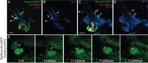
Anterior NeuroD:EGFP cells are migratory endocrine precursors. (A-D) Confocal sections of 3 days post fertilization (dpf) TgBAC(NeuroD:EGFP)nl1; Tg(ins:dsRed)m1018 embryos colabeled with antibodies against green fluorescent protein (GFP) and duct marker 2F11 (in blue). GFP+/2F11+ cells emerge from the duct (arrows), some with long projections typical of migrating cells (A, B, arrowhead). An individual cell that has separated from the duct is weakly DsRed positive (C, arrowhead). (E) Confocal image projections from a timelapse sequence of a 3 dpf TgBAC(NeuroD:EGFP)nl1;Tg(-8.0cldnb:lynEGFP)zf106 double transgenic embryo [see Additional file [8]. Membrane-linked enhanced green fluorescent protein (EGFP) in Tg(-8.0cldnb:lynEGFP)zf106 embryos delineates the gut epithelium (blue outline = extrapancreatic duct (EPD)) while robust cytoplasmic EGFP is present in NeuroD-expressing cells. Two NeuroD:EGFP cells (arrow) adjacent to the EPD migrate to the principal islet. All embryos are shown from ventral view, with the anterior to the left. Scale bar = 15 μM.
|
