
FIGURE
Fig. 3
- ID
- ZDB-FIG-111215-18
- Publication
- Nguyen et al., 2011 - A high level of liver-specific expression of oncogenic KrasV12 drives robust liver tumorigenesis in transgenic zebrafish
- Other Figures
- All Figure Page
- Back to All Figure Page
Fig. 3
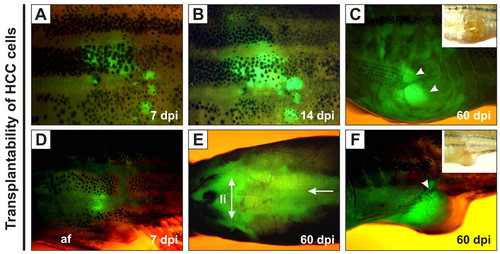
Growth of transplanted krasV12 liver tumors in WT recipients. (A–F) EGFP-positive HCC cells were transplanted intraperitoneally into irradiated recipients. (A,D) EGFP fluorescence was observed near the sites of injection at 7 dpi. af, anal fin. (B) Transplanted cells proliferated by 14 dpi. (E) Extensive infiltration of tumor cells along the peritoneal cavity (arrow), especially in the region surrounding the liver (li; double-headed arrow), was observed at 60 dpi. (C,F) The outgrowth of EGFP-positive tumor mass (arrowheads) penetrated into the abdominal wall and/or peritoneal cavity at 60 dpi. |
Expression Data
Expression Detail
Antibody Labeling
Phenotype Data
Phenotype Detail
Acknowledgments
This image is the copyrighted work of the attributed author or publisher, and
ZFIN has permission only to display this image to its users.
Additional permissions should be obtained from the applicable author or publisher of the image.
Full text @ Dis. Model. Mech.