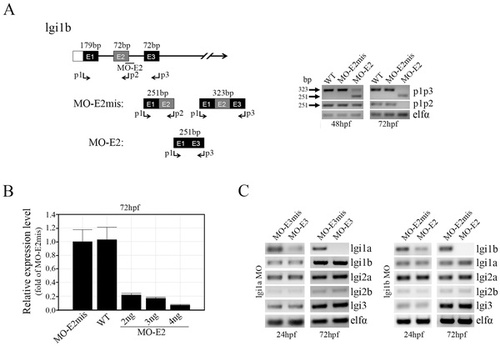
Splice-site-targeted morpholino oligonucleotides alter normal lgi1b splicing. (A) A schematic diagram of the partial pre-mRNA map showing the predicted outcomes in lgi1b morphants (left panel). The exons are shown in boxes, labeled with the corresponding exon number E1–E3, and the introns are represented by solid lines. The location of the exon 2 splice site, targeted by the MO-E2 morpholino, and the primer binding sites (p1, p2, p3, arrows) for PCR validation are indicated. Semi-qRT-PCR analysis (right panel) of lgi1b transcript levels in wild-type fish, MO-E2mis and MO-E2 morphants at different time points (48 and 72 hpf) show aberrant splicing in the MO-E2 (2 ng) morphants which generates an abnormal (251 bp) mRNA (lacking exon 2) using primers p1/p3. (B) qRT-PCR analysis of lgi1b transcript levels in wild-type, MO-E2mis and MO-E2 morphants following injection of three different concentrations of MO-E2 (2, 3 and 4 ng). (C) Semi-qRT-PCR analysis of transcript levels of the zebrafish lgi1 family members after knockdown of lgi1a (left) and lgi1b (right) respectively.
|
