
Fig. S2
- ID
- ZDB-FIG-111202-24
- Publication
- Feng et al., 2010 - T-lymphoblastic lymphoma cells express high levels of BCL2, S1P1, and ICAM1, leading to a blockade of tumor cell intravasation
- Other Figures
- All Figure Page
- Back to All Figure Page
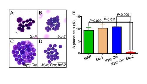
Bcl-2 Overexpression Inhibits Cell Proliferation in Myc-induced T-LBL in Zebrafish. (A-D) GFP-expressing thymocytes were FACS-sorted and stained with May-Grünwald-Giemsa to analyze the morphology of cells from (A) rag2-GFP (GFP), (B) rag2-EGFP-bcl-2 (bcl-2), (C) rag2-LDL-EGFP-mMyc;hsp70-Cre (Myc;Cre) and (D) rag2-LDL-EGFP-mMyc;hsp70-Cre;rag2-EGFP-bcl-2 (Myc;Cre;bcl-2) transgenic fish. (E) DNA flow cytometry demonstrates a decreased percentage of Myc;Cre;bcl-2 tumor cells in S phase, compared to control GFP, bcl-2, and Myc;Cre tumor cells (mean ± SD for GFP, bcl-2, Myc;Cre, Myc;Cre;bcl-2: 9.31 ± 2.39; 10.27 ± 3.29; 10.76 ± 0.54; 0.65 ± 0.61, respectively; n=4 fish per group). Scale bar for panels A-D = 10 μm. |
Reprinted from Cancer Cell, 18(4), Feng, H., Stachura, D.L., White, R.M., Gutierrez, A., Zhang, L., Sanda, T., Jette, C.A., Testa, J.R., Neuberg, D.S., Langenau, D.M., Kutok, J.L., Zon, L.I., Traver, D., Fleming, M.D., Kanki, J.P., and Look, A.T., T-lymphoblastic lymphoma cells express high levels of BCL2, S1P1, and ICAM1, leading to a blockade of tumor cell intravasation, 353-366, Copyright (2010) with permission from Elsevier. Full text @ Cancer Cell