
Fig. 4
- ID
- ZDB-FIG-111129-9
- Publication
- Alexander et al., 2011 - Combinatorial roles for BMPs and Endothelin 1 in patterning the dorsal-ventral axis of the craniofacial skeleton
- Other Figures
- All Figure Page
- Back to All Figure Page
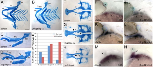
Exogenous BMP is sufficient to induce ventral arch identity. (A-D,F-H) Alcian Blue-stained cartilages of 4-5 dpf larvae, dissected and flat-mounted, anterior towards the left. (I-N) Whole-mount in situ hybridization at 30 hpf, lateral views, anterior towards the left. (A,B) Pharyngeal cartilages of a control (A) and an embryo implanted behind the eye at 20 hpf with a bead soaked in 20 μg/μl of human recombinant BMP4/7 heterodimers (B). (C,D) Isolated cartilages of the mandibular (1) and hyoid (2) arches: control (C); BMP4/7 bead-implanted (D). Grey arrowheads in B,D indicate duplicated Mc cartilages. (E) Histogram of phenotype frequency in bead-implanted embryos depending on BMP4/7 concentration. (F-H) Neurocranial cartilages: control (F); BMP4/7 bead-implanted (G); heat-shocked Tg(hsp70I:Gal4;UAS:Bmp4) (H). Black arrowheads indicate ectopic cartilages. (I,J) dlx6a expression slightly expands dorsally (arrow) in response to a BMP4/7-soaked bead (J). (K,L) dlx3b expression does not change when BMP4/7-soaked beads are implanted. (M,N) hand2 expression also expands dorsally (arrow) in response to BMP4/7 protein. Asterisks and blue circles indicate beads. 1, mandibular arch; 2, hyoid arch; Ch, ceratohyal; e, ethmoid plate; Hm, hyomandibular; Mc, Meckel’s cartilage; Mc’, ectopic Meckel’s cartilage; Pq, palatoquadrate; tr, trabeculae. Scale bars: 100 μm. |