
Fig. 1
- ID
- ZDB-FIG-111129-6
- Publication
- Alexander et al., 2011 - Combinatorial roles for BMPs and Endothelin 1 in patterning the dorsal-ventral axis of the craniofacial skeleton
- Other Figures
- All Figure Page
- Back to All Figure Page
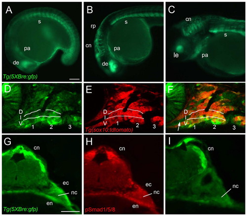
Expression of Tg(Bre:GFP) in the pharyngeal arches. (A-C) GFP fluorescence in living Tg(Bre:GFP) transgenic embryos, lateral views, anterior towards the left. (D-F) Lateral views of the arches at higher magnification. Confocal slices of Tg(Bre:GFP) (D) and Tg(sox10:lyn-tdTomato) (E) double transgenics. The two channels are merged in F, demonstrating direct BMP responses in NC and in the stomodeum (arrow). (G-I) Adjacent transverse sections through the hindbrain and arches stained with anti-GFP (G,I) and anti-pSmad1/5/8 (H), revealing BMP responses in the NC and in surrounding endoderm (en) and ectoderm (ec). (A) 16 hpf. (B) 24 hpf. (C) 48 hpf. (D-F) 28 hpf. (G-I) 30 hpf. cn, commissural neurons; D, dorsal; de, dorsal eye; ec, ectoderm; en, endoderm; I, intermediate; le, lens; nc, neural crest; pa, pharyngeal arches; rp, roofplate; s, somites; V, ventral. Scale bars: 100 μm. |