
Fig. 5
- ID
- ZDB-FIG-111129-5
- Publication
- Huang et al., 2011 - Phosphorylation of the Zebrafish M6Ab at Serine 263 Contributes to Filopodium Formation in PC12 Cells and Neurite Outgrowth in Zebrafish Embryos
- Other Figures
- All Figure Page
- Back to All Figure Page
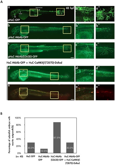
Expression of M6Ab-GFP and M6Ab(S263D)-GFP driven by a neuron-specific HuC promoter in zebrafish embryos. (A) To express the wild-type and S263D mutant proteins of zebrafish M6Ab under the control of a neuron-specific HuC promoter, plasmids pHuC-GFP, pHuC-M6Ab-GFP, pHuC-M6Ab(S263D)-GFP, and pHuC-M6Ab-GFP/pHuC-CaMKII · 1(T287D)-DsRed were individually injected into zebrafish embryos at the one-cell stage. Zebrafish embryos at 48 h post-fertilization (hpf) with GFP fluorescence were selected for the image analysis. Images were taken using a Zeiss LSM510 laser scanning confocal microscope. Merged images of red and green fluorescence are shown in (e, e2, and e3), while only green fluorescence images are shown in (a, a2, a3, b, b2, b3, c, c2, c3, d, d2 and d3). Higher magnification of two regions marked with yellow boxes in panel a-e is shown in panels a2–e2 and a3–e3, respectively. (B) Quantification of zebrafish numbers with significant neurite outgrowth at 48 hpf. Scale bars, 10 μm. |