|
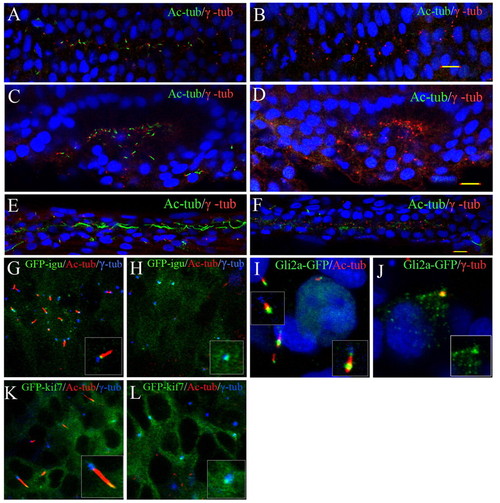
Disruption of primary ciliogenesis and aberrant localization of Hh signalling components in MZta3 embryos. (A-D) Confocal images of embryos fixed at the 10-somite (A,B) or 14-somite stage (C,D), and double stained with anti-acetylated tubulin (green) and anti-β tubulin (red) revealing the absence of cilia from the neural tube (B) and otic vesicle (D) of MZta3 mutants. (E,F) Similar preparations of embryos fixed at 24 hpf showing absence of long motile cilia from the pronephric duct of MZta3 mutants (F). (G-L) Localization of Hh pathway components in the primary cilia of somitic cells in 5- to 10-somite stage wild-type and MZta3 mutant embryos. Accumulation of a GFP-Dzip1 fusion protein in the basal body (G) is unaffected in MZta3 embryos (H). (I,K) GFP-tagged forms of Gli2a (I) and Kif7 (K) accumulate at the tip of primary cilia in wild-type cells; in the absence of the primary cilium in MZta3 embryos, both proteins shows a punctate localization in the basal body (J,L).
|