
Fig. 2
- ID
- ZDB-FIG-111117-24
- Publication
- Korzh et al., 2011 - The interaction of epithelial Ihha and mesenchymal Fgf10 in zebrafish esophageal and swimbladder development
- Other Figures
- All Figure Page
- Back to All Figure Page
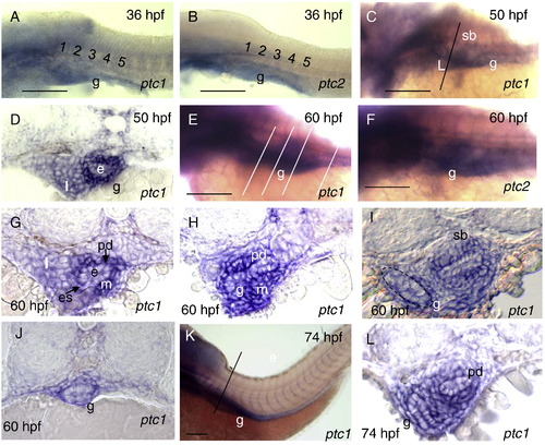
Expression pattern of ptc1 and ptc2 during development of the gut, pneumatic duct and swimbladder. A, B, Expression of ptc1 and ptc2 is detected by WISH at 36 hpf, C, ptc1 expression increased along the foregut and swimbladder bud at 50 hpf. A–C, Whole mount, lateral view. D, Cross-section of embryos in C revealed strong expression in the esophagus mesenchyme. E, F Expression of ptc1 and ptc2 is strong at 60 hpf. G–I, Cross-sections showed ptc1 expression domain in the mesenchyme surrounding epithelium of the esophagus, pneumatic duct, swimbladder and intestine. Note strong expression in the esophagus and along pneumatic duct. J, Cross section shows ptc1 expression in the posterior gut mesenchyme at 60 hpf. K, Expression of ptc1 at 74 hpf. L, Cross-section in K revealed strong expression of ptc1 in the mesenchyme surrounding the intestinal bulb, pneumatic duct and swimbladder. Abbreviations: e, epithelium; es, esophagus; g, gut, n, notochord; m, mesenchyme; pd, pneumatic duct; l, liver; sb, swimbladder. Scale bars: 250 μm. |
| Genes: | |
|---|---|
| Fish: | |
| Anatomical Terms: | |
| Stage Range: | Prim-25 to Protruding-mouth |
Reprinted from Developmental Biology, 359(2), Korzh, S., Winata, C.L., Zheng, W., Yang, S., Yin, A., Ingham, P., Korzh, V., and Gong, Z., The interaction of epithelial Ihha and mesenchymal Fgf10 in zebrafish esophageal and swimbladder development, 262-76, Copyright (2011) with permission from Elsevier. Full text @ Dev. Biol.