
Fig. S1
- ID
- ZDB-FIG-111031-8
- Publication
- Kroehne et al., 2011 - Regeneration of the adult zebrafish brain from neurogenic radial glia-type progenitors
- Other Figures
- All Figure Page
- Back to All Figure Page
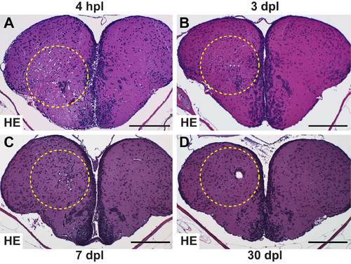
Timecourse of status spongiosus and re-establishment of tissue integrity. (A) At 4 hpl the majority of the cross-sectional area of the lesioned hemisphere has the typical sieve-like structure known as status spongiosus. In addition, tissue outside the stab canal shows a spongious phenotype due to oedema. (B) By 3 dpl, the vacuolated area is dramatically reduced in size compared with at 4 hpl (A). The size of the vacuoles is strongly reduced compared with 1 dpl (Fig. 1B). (C) The oedema size and the degree of vacuolisation is further decreased at 7 dpl compared with 3 dpl. (D) The oedema size is further reduced at 30 dpl. Often a single small hole is seen. Scale bars: 200 μm. All panels show HE stains of 1 μm paraffin sections. Dashed outlines represent the lesion canal. |