
Fig. 6
- ID
- ZDB-FIG-111031-6
- Publication
- Kroehne et al., 2011 - Regeneration of the adult zebrafish brain from neurogenic radial glia-type progenitors
- Other Figures
- All Figure Page
- Back to All Figure Page
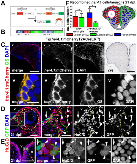
Newly generated neurons derive from a her4.1-expressing radial glia-type progenitor. (A) Generation of double-transgenic fish used for cell fate analysis: fish expressing CreERT2 controlled by the her4.1-promotor were crossed to red-to-green reporter fish. In double-transgenics, GFP expression can be specifically induced in her4.1-expressing radial glia and their progeny by tamoxifen application and heat shock. (B) Recombination was induced specifically in her4.1-expressing radial glia by tamoxifen injection 1 day before lesion. Fish (18 to 21 dpl) were heat-shocked to induce GFP expression in recombined cells only. (C) Cre-recombinase expression is restricted to radial glia in the adult telencephalon of Tg(her4.1:mCherryT2ACreERT2) fish: the expression of mCherry (red) is restricted to ventricular cells that resemble radial glia by morphology. Cre mRNA expression (blue) is restricted to the VZ, as shown by in situ hybridization. her4.1:mCherry is expressed in ventricular cells that co-express glutamine synthetase (GS, green), identifying them as radial glia. (D) By 21 dpl, many recombined GFP+(green)/HuC/D+(red) neurons/neuroblasts (arrowheads) are found within the lesion-canal (yellow outline) and in the PVZ (white outline). In the VZ recombined GFP+/HuC/D– cells with radial processes, i.e. radial glia (arrow), are found. (E) Recombined neurons (arrowheads) derived from her4.1+ radial glia (arrow) are exclusively found in the PVZ in the dorsal part of unlesioned control hemispheres 21 dpl. Scale bars: 200 μm in C overviews, inset 10 μm; 100 μm in D, inset 50 μm; 100 μm in E, inset 10 μm. v, ventricle; l, lesioned hemisphere; c, control hemisphere. (F) Quantification of cell fate analysis 21 dpl (n=7). The number of recombined GFP+/HuC/D– ventricular radial glia is not significantly different in lesioned and control hemispheres. However, the number of lineage-traced neurons is significantly increased in the lesioned hemisphere, showing that radial glia in the lesioned hemisphere produce more neurons and, in particular, give rise to neurons that migrated into the parenchyma. Data are mean+s.e.m. *Pd0.01. |