
Fig. 3
- ID
- ZDB-FIG-111028-14
- Publication
- Palencia-Desai et al., 2011 - Vascular endothelial and endocardial progenitors differentiate as cardiomyocytes in the absence of Etsrp/Etv2 function
- Other Figures
- All Figure Page
- Back to All Figure Page
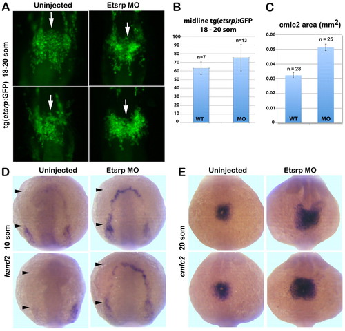
Migration of etsrp:GFP-expressing cells is not affected while myocardial markers are expanded in etsrp morphants. (A) etsrp:GFP expression in the midline population (white arrows) of presumptive endocardial progenitors is not affected in Etsrp morphants (right panels) at the 18- to 20-somite stages. Maximum intensity projection of fixed Tg(etsrp:GFP) embryos, ventral flat-mounted view, anterior is upwards. (B) At the 18- to 20-somite stages, the relative numbers of etsrp:GFP+ putative cardiac progenitors that migrate to the midline are similar between uninjected controls and etsrp morphants. Data are mean±s.e.m. (C) The calculated average area (mm2) of cmlc2 in situ hybridization staining at the 20-somite stage shows a 58% increase in etsrp morphants compared with uninjected wild-type controls depicted in E. (D) hand2 expression extends into the rostral ALPM (black arrowheads) in etsrp morphants but is absent from this region in the uninjected controls at the 10-somite stage. (E) cmlc2 expression at 20-somite stage reveals radial expansion of the cardiac plate in etsrp morphants (right panels) compared with uninjected wild-type embryos (left panels). Anterodorsal whole-mount view, anterior is upwards. |