
Fig. S4
- ID
- ZDB-FIG-111017-12
- Publication
- Ramachandran et al., 2011 - Ascl1a/Dkk/ beta -catenin signaling pathway is necessary and glycogen synthase kinase-3 beta inhibition is sufficient for zebrafish retina regeneration
- Other Figures
- All Figure Page
- Back to All Figure Page
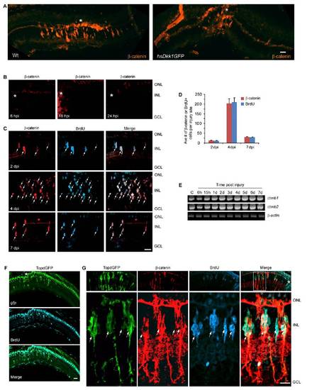
β-catenin stabilization in the injured retina. (A) At 4 dpi epitope retrieval reveals β-catenin stabilization localized to the injury site (asterisk) in Wt fish. Overexpression of Dkk1b in hsDkk1GFP transgenic fish suppressed proliferation of MG-derived progenitors in the injured retina. (B) Epitope retrieval shows β-catenin is undetectable at 6–24 hpi. (C) Epitope retrieval shows β-catenin is first detected at 2 dpi in proliferating MG-derived progenitors (BrdU+). (D) Quantification of β-catenin+ and BrdU+ cells in the injured retina. (E) The two different β-catenin mRNA isoforms, ctnnb1 and ctnnb2, are expressed in the uninjured and injured retina and show marginal increases following injury. (F) Low magnification view of retinal section in TOPdGFP transgenic fish showing transgene gfp expression localized to the injury site (in situ hybridization) overlaps with BrdU+ cells (immunofluorescence). (G) At 4 dpi retinal progenitors (BrdU+) coexpress β-catenin and GFP in TOPdGFP transgenic fish. Upper shows a low magnification view of the retina, and Lower shows a high magnification view of the boxed area in the low magnification merge panel. Asterisks indicate injury sites. (Scale bar, 10 μm.) ONL, outer nuclear layer; INL, inner nuclear layer; GCL, ganglion cell layer. |