
Fig. 2
- ID
- ZDB-FIG-111013-9
- Publication
- Waskiewicz et al., 2001 - Zebrafish Meis functions to stabilize Pbx proteins and regulate hindbrain patterning
- Other Figures
- All Figure Page
- Back to All Figure Page
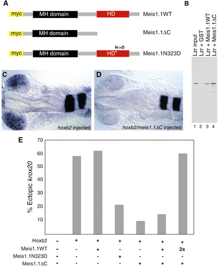
Expression of dominant-negative Meis1.1 inhibits Hoxb2 function. (A) Meis1.1WT was mutated in two alternative ways to generate forms that can bind Lzr but cannot bind DNA, Meis1.1ΔC and Meis1.1N323D. (B) To confirm that deletion of the Meis C terminus does not inhibit Pbx binding and to demonstrate that Meis1.1 can bind Lzr, the proteins were synthesized in vitro and assayed for ability to bind one another. Lane 1 contains 5% of the input Lzr, while lanes 2, 3 and 4 display proteins that bind to GST (lane 2), GST-Meis1.1 (lane 3), or GST-Meis1.1ΔC (lane 4). Binding between Lzr and Meis proteins varies from 10%-30% depending on the stringency of the wash conditions (data not shown). (C,D) hoxb2 overexpression results in ectopic expression of krox20 within the retina of approximately 60% of injected embryos. (C) 60% embryos have expression of retinal krox20 shown here at 20 somites. (D) 90% of embryos injected with hoxb2 and meis1.1ΔC contain undetectable levels of ectopic krox20. (E) Quantification of embryos expressing ectopic krox20 in the eye after injection of hoxb2 and dominant-negative meis RNAs. |