
Fig. 8
- ID
- ZDB-FIG-111013-15
- Publication
- Waskiewicz et al., 2001 - Zebrafish Meis functions to stabilize Pbx proteins and regulate hindbrain patterning
- Other Figures
- All Figure Page
- Back to All Figure Page
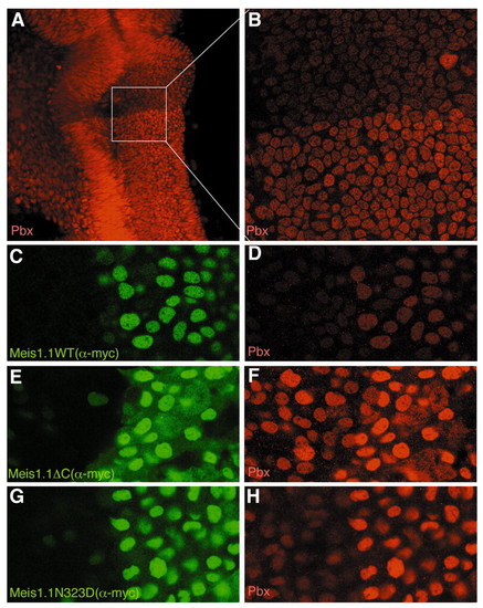
Meis stabilizes endogenous nuclear Pbx protein. (A) 24 hour embryo stained with pan-Pbx antiserum, showing a prominent boundary of nuclear staining at the r1/r2 boundary with higher Pbx levels posterior to the boundary. (B) Higher magnification image of box outlined in A, demonstrating that Pbx proteins are predominantly nuclear on both sides of the boundary. (C-H) Two to four somite stage embryos expressing Meis1.1WT (C,D), Meis1.1ΔC (E,F) and Meis1.1N323D (G,H), stained with 9E10 to visualize the Myc epitope on the Meis proteins (green staining in C,E,G) and with α-pan-Pbx antibody to detect endogenous Pbx protein (red staining in D,F,H). Note that cells expressing Meis1.1WT or mutant protein exhibit stronger Pbx immunoreactivity, whereas an unrelated Myc-tagged protein did not have this effect (data not shown). Also note that all Meis forms are predominantly nuclear. |