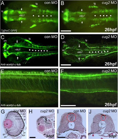
cug2 deficiency causes neurodegeneration in developing embryos. A, B. In huC:GFP transgenic embryos, injection of cug2 MO (B) causes a reduction in the number of neurons in the nucleus of the medial longitudinal fasciculus (nMLF, arrows) and the rhombomere (white spots) compared to control MO (A). Scale bar = 200 μm. C, D. Anti-acetylated α-tubulin staining of the brain of control (C) and cug2 morphants (D). cug2 deficiency causes axonal scaffolding defects in the anterior commissure (ac), olfactory nerve (ofn), nMLF (arrows), and hindbrain commissure (white spots) at 26 hpf. Scale bar = 200 μm. E, F. Anti-acetylated α-tubulin staining of the spinal cord of cug2 morphants (F) at 26 hpf shows reduced arborization in Rohon-Beard (RB) sensory neurons compared to control (E). Scale bar = 100 μm. G-J. Histological sections of control (G, I) and cug2 MO (H, J)-injected embryos at 3 dpf. cug2 morphants exhibit severely disrupted retina layer formation (H) and a much smaller neural tube that contains fewer cells (J). gcl, ganglion cell layer; inl, inner nuclear layer; ipl, inner plexiform layer; le, lens; n, notochord; on, optic nerve; opl, outer plexiform layer; pcl, photoreceptor cell layer; pe, pigmented epithelium. Scale bars = 50 μm.
|
