
Fig. 2
- ID
- ZDB-FIG-110929-49
- Publication
- Wythe et al., 2011 - Hadp1, a newly identified pleckstrin homology domain protein, is required for cardiac contractility in zebrafish
- Other Figures
- All Figure Page
- Back to All Figure Page
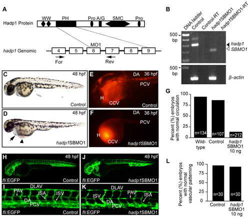
Knockdown of hadp1 decreases cardiac output and contractility in zebrafish. (A,B) Schematic representation of Hadp1 protein (top) and partial genomic structures (bottom; boxes represent coding exons), with positions of the MO and RT-PCR primers indicated. See Fig. 1 for abbreviations. (B) RT-PCR analysis of control and morphant embryos. β-actin is a loading control. –RT indicates the absence of reverse transcriptase as a negative control. (C,D) Brightfield images of live embryos (anterior is to the left) after injection with 10 ng control MO or 10 ng SBMO1. Arrow and arrowhead indicate pericardial effusion and blood pooling, respectively. (E,F) Microangiography following injection of control MO or SBMO1 reveals that circulation is altered following knockdown of hadp1. Heart (H), common cardinal vein (CCV), dorsal aorta (DA) and the posterior cardinal vein (PCV) are indicated. Circulation was scored by microangiography, or under transmitted light, and the results are quantified in G. (H–K) Confocal micrographs of control MO or SBMO1 morphant fli:EGFP transgenic embryos. Dorsal aorta (DA), posterior cardinal vein (PCV), parachordal vein (PAV), intersegmental arteries (ISA), intersegmental vessel (ISV) and dorsal longitudinal anastomotic vessel (DLAV) are indicated. (L) Quantification of normal vascular patterning. |
| Fish: | |
|---|---|
| Knockdown Reagent: | |
| Observed In: | |
| Stage Range: | Prim-25 to Long-pec |