
Fig. 5
- ID
- ZDB-FIG-110929-47
- Publication
- Jayasena et al., 2011 - Live imaging of endogenous Collapsin response mediator protein-1 expression at subcellular resolution during zebrafish nervous system development
- Other Figures
- All Figure Page
- Back to All Figure Page
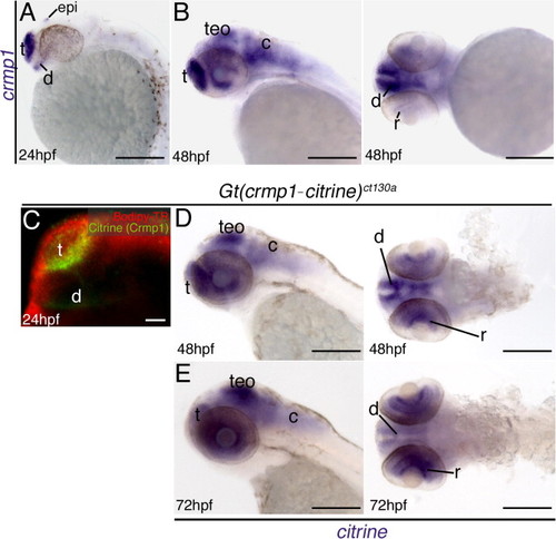
In situ hybridization results showing expression of crmp1 (A)–(B) and citrine (D)–(E). In wild type fish crmp1 expression is localized to the nervous system at 24 hpf (A) and 48 hpf (B). (B) Lateral and frontal views at 48 hpf. (C) Confocal z-stack at 24 h post fertilization (hpf) showing Citrine/ Crmp1 (green) expression in the forebrain regions. Red: Bodipy-TR vital dye. (D)–(E) Lateral and ventral views at 48 hpf and 72 hpf. Transcripts for citrine are expressed in selected neural regions at both stages consistent with wild type expression. Abbreviations: epi, epiphysis; t, telencephalon; teO, optic tectum/midbrain; c, cerebellum/ hindbrain; d, diencephalon; r, retina. Scale: 200 μM, 50 μM (C). |
| Genes: | |
|---|---|
| Fish: | |
| Anatomical Terms: | |
| Stage Range: | Prim-5 to Protruding-mouth |
Reprinted from Gene expression patterns : GEP, 11(7), Jayasena, C.S., and Bronner-Fraser, M., Live imaging of endogenous Collapsin response mediator protein-1 expression at subcellular resolution during zebrafish nervous system development, 395-400, Copyright (2011) with permission from Elsevier. Full text @ Gene Expr. Patterns