
Fig. 2
- ID
- ZDB-FIG-110920-90
- Publication
- Smith et al., 2011 - Transmembrane protein 2 (Tmem2) is required to regionally restrict atrioventricular canal boundary and endocardial cushion development
- Other Figures
- All Figure Page
- Back to All Figure Page
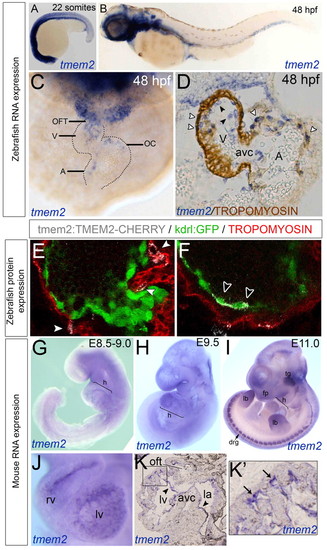
tmem2 expression in the endocardial cells of zebrafish and mouse hearts and in the myocardial cells of zebrafish hearts. (A-C) ISH for zebrafish tmem2 expression at 22 somites (A) and 48 hpf (B,C) shown in lateral (A,B) or frontal (C) view. (D) Transverse section of tmem2 ISH with tropomyosin counterstaining. White arrowheads, myocardial tmem2 expression; black arrowheads, endocardial tmem2 expression. (E,F) Transient expression from a Tmem2-Cherry BAC construct. White arrowheads, myocardial Tmem2-Cherry; black arrowheads, endocardial Tmem2-Cherry. (G-K′) ISH of mouse embryos stained for Tmem2 at E8.5-9.0 (G), E9.5 (J-K′) and E11.0 (I). (K) Sagittal sectioning revealed expression in the endocardium (arrows). (K2) Enlargement of boxed region in K. Arrows indicate staining in the ventricular trabeculae. A, atrium; avc, atrioventricular canal; drg, dorsal root ganglion; fp, facial prominences; h, heart; la, left atrium; lb, limb buds; lv, left ventricle; OC, outer curvature; OFT, outflow tract; tg, trigeminal ganglion; V, ventricle. |
| Gene: | |
|---|---|
| Fish: | |
| Anatomical Terms: | |
| Stage Range: | 20-25 somites to Long-pec |