
FIGURE
Fig. S1
- ID
- ZDB-FIG-110909-8
- Publication
- Taylor et al., 2011 - Differentiated melanocyte cell division occurs in vivo and is promoted by mutations in Mitf
- Other Figures
- All Figure Page
- Back to All Figure Page
Fig. S1
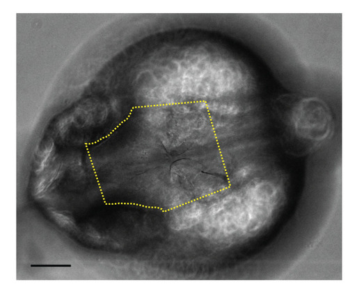
Melanocyte counting. All melanocyte counts took place on a defined head region, which extended from halfway between the eyes down to ears (yellow dotted line). Scale bar: 100 microns. |
Expression Data
Expression Detail
Antibody Labeling
Phenotype Data
Phenotype Detail
Acknowledgments
This image is the copyrighted work of the attributed author or publisher, and
ZFIN has permission only to display this image to its users.
Additional permissions should be obtained from the applicable author or publisher of the image.
Full text @ Development