
Fig. 7
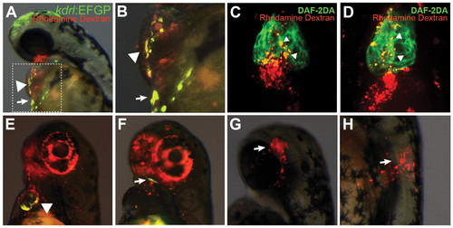
Cells overexpressing gata5/smarcd3b form multiple cardiovascular lineages. (A,B) Donor kdrl:EGFP embryos were injected at the one-cell stage with gata5/smarcd3b RNA and tetramethylrhodamine dextran, and transplanted to the animal pole of wild-type host embryos at 4 hpf. Contribution to endocardium (arrowheads) and blood vessels neighboring the heart (arrows) was evident at 48 hpf. Lateral view of a 48 hpf embryo is shown, with the boxed region in A shown at higher magnification in B. (C,D) Donor embryos bearing no transgene were injected with gata5/smarcd3b RNA and tetramethylrhodamine dextran, and transplanted as in A,B. At 72 hpf, DAF-2DA staining was carried out. Arrowheads indicate the overlap of DAF-2DA staining (green) and tetramethylrhodamine dextran (red) in the outflow tract of the heart. Ventral view with anterior towards the top of images. (E,F) Donor Tg(tcf21:GVEcR;UAS:EGFP) embryos were injected with gata5/smarcd3b RNA and tetramethylrhodamine dextran and transplanted as in A,B. Contribution to ventricle (E, arrowhead) and head mesoderm/facial muscle (F, arrow) was evident at 48 hpf. (G,H) Heterochronic transplantation of 4 hpf donor cells from myl7:EGFP embryos injected with gata5/smarcd3b RNA and tetramethylrhodamine dextran to the head (G) and trunk (H) of 16 hpf host embryos. Combined bright-field/EGFP/rhodamine images are shown of transplant embryos at 48 hpf. Lateral views with anterior towards the top. |