
FIGURE
Fig. 8
- ID
- ZDB-FIG-110812-24
- Publication
- Liu et al., 2011 - Cell adhesion molecule cadherin-6 function in zebrafish cranial and lateral line ganglia development
- Other Figures
- All Figure Page
- Back to All Figure Page
Fig. 8
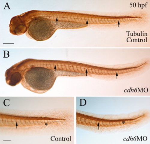
Normal development of the posterior lateral line nerve in cdh6 morphants. All panels show lateral views of whole-mount embryos (anterior to the left and dorsal up) processed for anti-acetylated tubuline immunoperoxidase staining. Arrows point to the posterior lateral line nerve, while the asterisk indicates the terminus of the nerve. C and D are higher magnifications (same magnification) of the tail region of the embryos in A and B (same magnification), respectively. Scale bar = 200 μm in A,B, 100 μm in C,D. |
Expression Data
Expression Detail
Antibody Labeling
Phenotype Data
Phenotype Detail
Acknowledgments
This image is the copyrighted work of the attributed author or publisher, and
ZFIN has permission only to display this image to its users.
Additional permissions should be obtained from the applicable author or publisher of the image.
Full text @ Dev. Dyn.