
Fig. 7
- ID
- ZDB-FIG-110812-23
- Publication
- Liu et al., 2011 - Cell adhesion molecule cadherin-6 function in zebrafish cranial and lateral line ganglia development
- Other Figures
- All Figure Page
- Back to All Figure Page
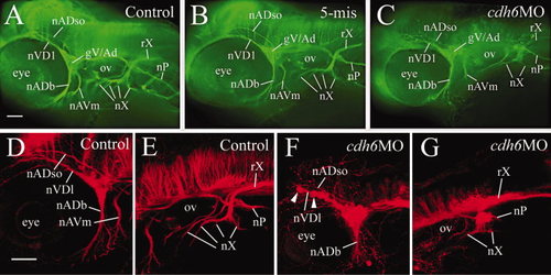
cdh6 loss-of-function defects in cranial and lateral line nerves, demonstrated by anti-acetylated tubulin immunofluorescent staining, in a control embryo (A), an embryo injected with the 5-mismatched MO (B), and a cdh6 morphant (C). The images are lateral views (anterior to the left and dorsal up) of the head region of the whole-mount embryos. D–G: Laser scanning confocal microscopy image projections confirmed results using wide field microscopy: anterior cranial nerves (D,F) and posterior cranial nerves (E,G) in control (D,E) and cdh6 morphant (F,G) embryos. Arrowheads in F indicate two cell clusters of the fragmented gV/Ad. The morphant vagus root (rX, G) was difficult to discern because it was closely opposed to anti-acetylated tubulin-positive brain cells and fiber tracks. Other abbreviations: nADb, buccal ramus of the anterodorsal lateral line nerve; nADso, superior ophthalmic ramus of the anterodorsal lateral line nerve; nAVm, mandibular ramus of the anteroventral lateral line nerve; nVDl, dorsolateral nerve of the trigeminal ganglion; nP, posterior lateral line nerve; nX, vagus nerves; ov, otic vesicle. All images have the same magnification. Scale bars = 50 μm. |
| Fish: | |
|---|---|
| Knockdown Reagent: | |
| Observed In: | |
| Stage: | Long-pec |