
Fig. 4
- ID
- ZDB-FIG-110719-23
- Publication
- Muto et al., 2011 - Genetic visualization with an improved GCaMP calcium indicator reveals spatiotemporal activation of the spinal motor neurons in zebrafish
- Other Figures
- All Figure Page
- Back to All Figure Page
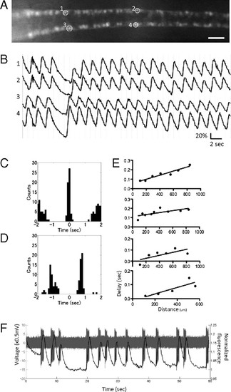
Spatiotemporal patterns of activation of the CaP motor neurons. (A) A dorsal view of the SAIGFF213A;UAS:GCaMPHS4A embryo at 24 hpf treated with 100 μM D-tubocurarine. (Scale bar, 200 μm.) (B) The fluorescence changes in ROIs in A. (C) Cross-correlogram of the ipsilateral CaP neurons. (D) Cross-correlogram of the contralateral CaP neurons. (E) The averaged timing of the peaks of the ipsilateral CaP neurons located in a rostral (Left)-to-caudal (Right) sequence. Data from four embryos are shown. (F) A whole cell-patch recording and simultaneous calcium imaging of the CaP neurons during spontaneous contractions. Action potentials (gray) and calcium signals (black) are shown. |