
Fig. S3
- ID
- ZDB-FIG-110706-15
- Publication
- Chou et al., 2011 - Fascin 2b Is a Component of Stereocilia that Lengthens Actin-Based Protrusions
- Other Figures
- All Figure Page
- Back to All Figure Page
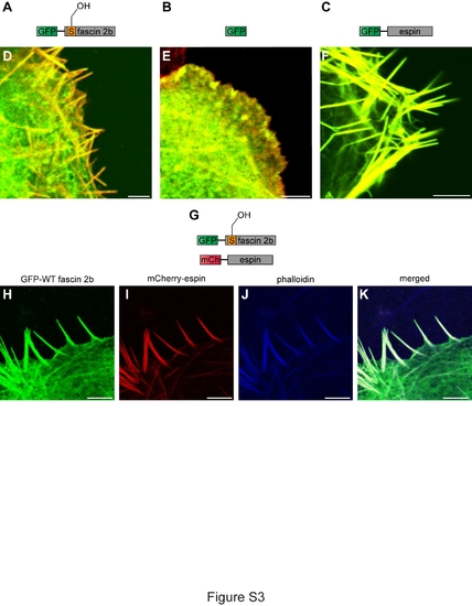
Localization of fascin 2b and espin proteins in fixed COS-7 cells. Schematics of the proteins expressed in cells are portrayed: GFP-WT fascin 2b (A), GFP (B), GFP-espin (C), and GFP-WT fascin 2b and mCherry-espin (G). To visualize actin-based filopodia, cells were labeled with Alexa 568 phalloidin (red)(D-F) or Alexa 633 phalloidin (blue) (J,K). Merged images show localization of GFP-WT fascin 2b (green) (D) or GFP-espin (green) (F) to filopodia where they overlap with actin (yellow). No filopodia are detectable in a fixed cell that expresses only GFP (green) (E). Coexpression of GFP-WT fascin 2b (H) and mCherry-espin (I) shows that both proteins colocalize (white) (K) to phalloidin-labeled filopodia (J) in a fixed COS-7 cell. Scale bars are 5 μm. |