
Fig. 1
- ID
- ZDB-FIG-110624-1
- Publication
- Row et al., 2011 - Completion of the epithelial to mesenchymal transition in zebrafish mesoderm requires Spadetail
- Other Figures
- All Figure Page
- Back to All Figure Page
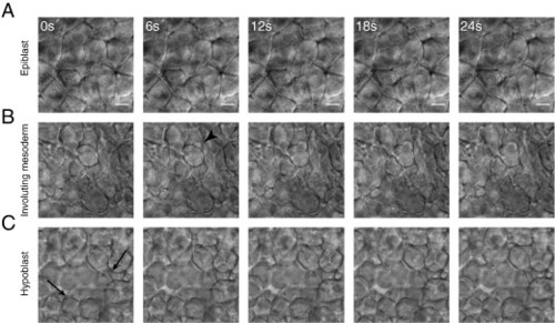
Cell morphology changes during mesoderm maturation. Cell morphology was observed using time-lapse DIC microscopy of the lateral margin during early gastrulation. (A) Cells in the outer (epiblast) layer did not extend protrusions and maintained long, stable, edge-to-edge contacts with their neighbors. (B) As cells underwent involution at the margin they remained tightly packed but extended many protrusions, primarily blebs (arrowhead). (C) Cells in the hypoblast displayed a mesenchymal morphology. They extended actin-based protrusions (arrows) as well as blebs, did not pack tightly, and formed only transient adhesions at discrete points of contact. All images are at the same magnification, scale bar is 10 μM. |
Reprinted from Developmental Biology, 354(1), Row, R.H., Maître, J.L., Martin, B.L., Stockinger, P., Heisenberg, C.P., and Kimelman, D., Completion of the epithelial to mesenchymal transition in zebrafish mesoderm requires Spadetail, 102-110, Copyright (2011) with permission from Elsevier. Full text @ Dev. Biol.