
Fig. 8
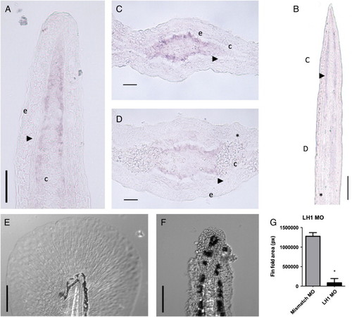
lh1 is implicated in actinotrichia synthesis. (A–D) In situ hybridization of lh1 in fin sections. Longitudinal sections of ray blastema at 3 (A) and 7 dpa (B). (C–D) Transversal sections of a 7-day regenerated fin at the positions outlined in (B). Arrowhead is actinotrichia. Asterisk is lepidotrichia. e is epidermis. c is connective tissue. (E–F) Morpholino knockdown of lh1 at 3 dpf (F) and 5 mismatch morphant control at 3 dpf (E). Fin fold areas (in pixels) are statistically compared following by Mann–Whitney U-test (G). Asterisk represents significant difference. Bars represent 25 (C and D) or 50 μm (A and B). |
| Fish: | |
|---|---|
| Knockdown Reagent: | |
| Observed In: | |
| Stage: | Protruding-mouth |
Reprinted from Developmental Biology, 354(1), Durán, I., Marí-Beffa, M., Santamaría, J.A., Becerra, J., and Santos-Ruiz, L., Actinotrichia collagens and their role in fin formation, 160-172, Copyright (2011) with permission from Elsevier. Full text @ Dev. Biol.