
Fig. 1
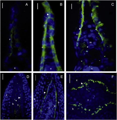
Actinotrichia formation during fin development and regeneration. Immunofluorescence with JAS′96 antibody against actinotrichia and AFCs (green) and hoechst nuclei staining (blue). (A–C) Fin fold sections at 30 (A), 36 (B) and 72 hpf (C). (D–E) Longitudinal sections of regenerating fin at 2 dpa (D) and 3 dpa (E). Transversal section at 5 dpa (F). Bars represent 10 (A–C) and 25 μm (D–F). Arrowhead is actinotrichia. Arrow is actinotrichia forming cell (AFC). e is epidermis. b is basal epidermal layer. c is connective tissue. n is notochord. Connective tissue and basal epidermal layer are separated by white lines in F. |
| Antibody: | |
|---|---|
| Fish: | |
| Condition: | |
| Anatomical Term: | |
| Stage Range: | Prim-5 to Day 5 |
Reprinted from Developmental Biology, 354(1), Durán, I., Marí-Beffa, M., Santamaría, J.A., Becerra, J., and Santos-Ruiz, L., Actinotrichia collagens and their role in fin formation, 160-172, Copyright (2011) with permission from Elsevier. Full text @ Dev. Biol.