
Fig. 7
- ID
- ZDB-FIG-110622-18
- Publication
- Alibaud et al., 2011 - A Mycobacterium marinum TesA mutant defective for major cell wall associated lipids is highly attenuated in Dictyostelium discoideum and zebrafish embryos
- Other Figures
- All Figure Page
- Back to All Figure Page
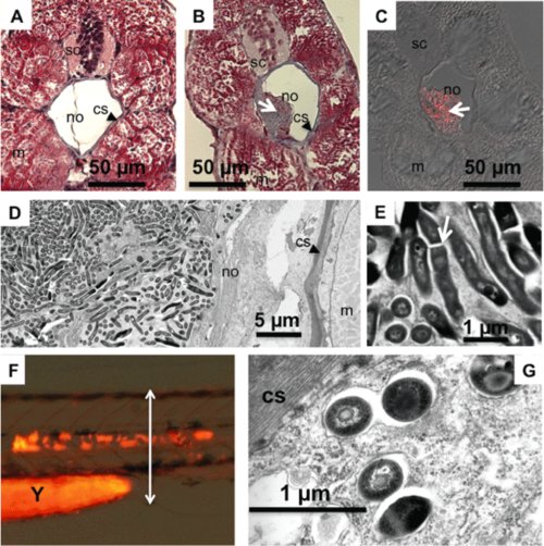
Histological and ultrastructural analyses of the tesA::Tn-infected notochord. A. Masson′s trichrome staining of a histological section from a non-infected notochord at 6 dpi. B. Same from a Mma tesA::Tn-infected notochord. C. TB Auramine-Rhodamine T staining of the section next to that in (B). Arrows indicate the focus of infection in (B) and (C). D, E and G. Transmission electron micrographs on an infectious focus in the infected notochord: (D) and (E) at 6 dpi; the arrow indicates a septum of division; (G) at 1 dpi. sc, spinal cord; m, muscular tissue; cs, collagen sheath surrounding the notochord. F. Micrograph at 1 dpi of the tesA::Tn_mCherry notochord infected embryo used for (G). The section displayed in (G) is depicted by the double-headed arrow. Magnification: ×10 000. Y, yolk. |