
Fig. s3
- ID
- ZDB-FIG-110622-128
- Publication
- Lupo et al., 2011 - Retinoic acid receptor signaling regulates choroid fissure closure through independent mechanisms in the ventral optic cup and periocular mesenchyme
- Other Figures
- All Figure Page
- Back to All Figure Page
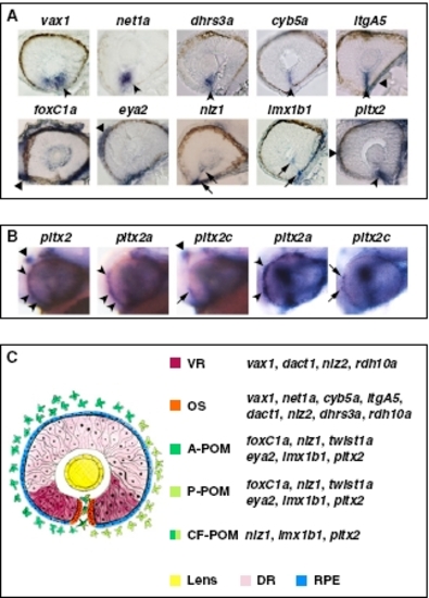
Expression patterns of VR/OS and POM genes. (A) Histological sections of 32- to 36-hpf eyes hybridized with the indicated probes. In the upper row, arrowheads point to expression in VR/OS cells, and the triangle points to itgA5 in extraocular tissue. In the lower row, triangles point to expression in POM around the optic cup, arrows point to expression in POM cells inside the choroid fissure, and the arrowhead points to pitx2 expression at the level of the choroid fissure, which might include OS cells besides POM. (B) Lateral views of 31-hpf heads (left three images) or eyes (right two images) hybridized with a pitx2 pan-isoform probe or with pitx2a- or pitx2c-specific probes, showing that pitx2a is the predominant isoform in the POM (arrowheads), whereas pitx2c is selectively expressed in the dorsal diencephalon (triangles). Arrows point to limited expression of pitx2c in few POM cells. (C) Schematic representation of the expression domains of VR/OS and POM genes, based on the data shown in A, Figs. 2 B and C and 3 B and C, and Fig. S5 A and B. A-POM, anterior POM; P-POM, posterior POM; CF-POM, POM located within the choroid fissure; DR, dorsal retina; RPE, retinal pigmented epithelium. |