
Fig. S4
- ID
- ZDB-FIG-110527-8
- Publication
- Krueger et al., 2011 - Flt1 acts as a negative regulator of tip cell formation and branching morphogenesis in the zebrafish embryo
- Other Figures
- All Figure Page
- Back to All Figure Page
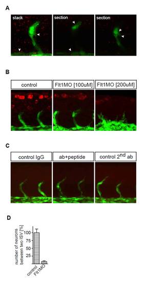
Flt1 immunohistochemistry. (A) Flt1 immunostaining (red) with tyramide signal amplification (TSA) in Tg(fli:egfp)y1 embryos. Note Flt1 immunostaining in the vessel and somite. (B) Flt1 immunostaining (red) in Tg(fli:egfp)y1 embryos in controls (left), after 3 ng flt1 ATG MO (middle) and after 6 ng flt1 ATG MO (right). Note the reduced immunostaining after MO-mediated knockdown of flt1. (C) Negative controls for immunostaining with the custom-made antibody against zebrafish Flt1 protein. Background staining was low in all three negative control immunostaining conditions. (Left) IgG, primary antibody (Ab) was against rabbit IgG. (Middle) Flt1Ab+peptide, embryos were incubated with primary antibody against Flt1 and the protein epitope used to generate the zebrafish Flt1 antibody prior to performing immunostaining. (Right panel) 2nd Ab, primary antibody omitted and stained with secondary antibody only. (D) Quantification of neuronal numbers in Tg(flt1BAC:yfp) × Tg(kdrl:ras-cherry) controls and flt1 morphants (percentage of control). Embryos were evaluated at 48 hpf. Error bars indicate s.e.m. |