
Fig. S2
- ID
- ZDB-FIG-110527-6
- Publication
- Krueger et al., 2011 - Flt1 acts as a negative regulator of tip cell formation and branching morphogenesis in the zebrafish embryo
- Other Figures
- All Figure Page
- Back to All Figure Page
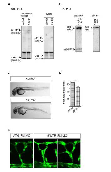
Comparison of ATG and 52 UTR targeting morpholinos, and western blot quantification of Flt1 protein. (A) Western blot analysis of zebrafish embryos using the Flt1 custom-made antibody after in vivo overexpression of mRNA encoding zebrafish mflt1 (left) and sflt1 (right). Note the clear increase in mFlt1 protein and sFlt1 protein after overexpression. (B) Immunoprecipitation analysis of purified GFP-mFlt1 fusion protein after overexpression in HeLa cells. GFP-mFlt1, GFP-zebrafish membrane-bound Flt1 fusion protein; GFP HC, GFP heavy chain. Left lane, detection of GFP signal; right lane, detection of Flt1 protein with custom-made antibody. Note that the custom-made Flt1 antibody detects the zebrafish Flt1 fusion protein. (C) Light microscopy images of control MO-injected embryo and embryo injected with flt1 ATG-targeting MO. (D) Heart rate was higher in control than in flt1 morphants. *P<0.05; Student′s t-test. Error bars indicate s.e.m. (E) Comparison of segmental branching after knockdown of flt1 with the ATG-targeting MO (left) and the 5′UTR-targeting MO (middle and right). Note that both the ATG- and the 5′UTR-targeting MO induced hyperbranching of the segmental artery sprouts. |