
Fig. 3
- ID
- ZDB-FIG-110527-19
- Publication
- Schnabel et al., 2011 - Regeneration of cryoinjury induced necrotic heart lesions in zebrafish is associated with epicardial activation and cardiomyocyte proliferation
- Other Figures
- All Figure Page
- Back to All Figure Page
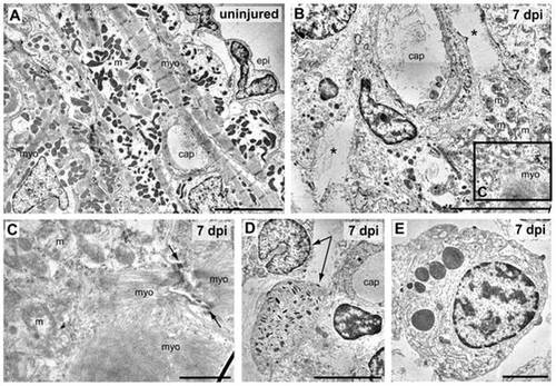
Ultrastructural analysis of cryolesioned myocardium reveals cardiomyocyte cell death and an inflammatory response. Electron micrographs of uninjured (A) and cryolesioned (7 dpi, B–E) zebrafish heart tissue. A normal organization of ventricular periphery with epicardal epithelium (epi) and subepicardial cardiomyocytes displaying well-organized striated myofilaments (myo) with Z lines and groups of electron dense mitochondria (m). A capillary (cap) is visible as well. (B) At 7 dpi, the lesioned area displays cellular debris and large tissue gaps (*) around a small capillary. In the lower right corner: cardiomyocyte with damaged mitochondria (m) and disorganised myofilaments (myo). (C) Magnification of the boxed area in B. A cryoinjured cardiomyocyte with disorganised mitochondria (m), myofilaments, and a loosened/ill defined intercalated disc (arrows) is shown. (D, E) Granulocytes in the wound area. (D) Heterophil granulocytes (arrows), the upper one with a peripheral, nonsegmented nucleus, the lower one with characteristic, cigar-shaped cytoplasmic granules. (E) Eosinophil granulocyte. Scale bars: 5 μm in A, B, and D, 1 μm in C, and 2 μm in E. |