|
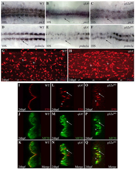
Convergence of qkA and Hh phenotypes. (A-F) qkAt and gli2aMO zebrafish embryos exhibit downregulated Hh signaling. Embryos were processed at the 10-somite stage for in situ hybridization with the indicated probes. Dorsal view. (G,H) Lateral views of Pax7-immunostained embryos at 24 hpf. Two populations of Pax7-positive cells can be discerned, of which dermomyotomal cells (arrowheads) exhibit the weaker expression. The number of dermomyotomal cells per somite is indicated beneath each somite. (I-Q) Slow fiber distal migration requires QkA and Hh activities. Embryos were stained at 24-hpf for slow fibers (F59) and all differentiated muscle fibers (MF20) as indicated, then analyzed by confocal microscopy (transverse sections). Note the presence of gaps (asterisks) and deep slow muscle cells (arrows) medial to the fast muscle cells in qkAt mutants and gli2a morphants.
|