
Fig. 3
- ID
- ZDB-FIG-110428-12
- Publication
- Bussmann et al., 2011 - Arterial-venous network formation during brain vascularization involves hemodynamic regulation of chemokine signaling
- Other Figures
- All Figure Page
- Back to All Figure Page
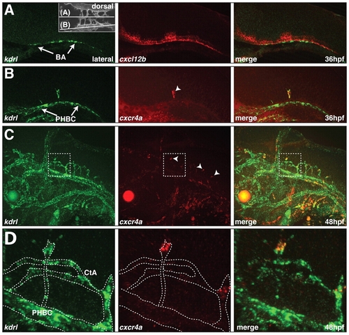
Expression of cxcl12b and cxcr4a in the zebrafish hindbrain. (A-D) Two-color fluorescent in situ hybridization showing the distribution of kdrl mRNA (green) and cxcl12b (A) and cxcr4a (B-D) mRNA (red). Lateral views, anterior to the left. (A) cxcl12b and kdrl expression at 36 hpf. Inset indicates the plane of view in A and B. (B) cxcr4a and kdrl expression at 36 hpf. Arrowhead indicates a single cxcr4a-expressing CtA sprout. (C) cxcr4a and kdrl expression at 48 hpf. Arrowheads indicate tip cells of CtA sprouts expressing cxcr4a. (D) Enlargement of the boxed area in C. The kdrl-positive endothelial cells are outlined. |
| Genes: | |
|---|---|
| Fish: | |
| Anatomical Terms: | |
| Stage Range: | Prim-25 to Long-pec |