
Fig. 3
- ID
- ZDB-FIG-110425-3
- Publication
- Herpin et al., 2007 - Specification of primordial germ cells in medaka (Oryzias latipes)
- Other Figures
- All Figure Page
- Back to All Figure Page
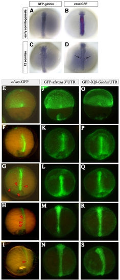
Stabilization of zebrafish vasa and translational regulation of medaka olvas mRNA in PGCs of medaka. 100 pg of GFP tagged fusion RNAs were injected at the 2-cell stage. (A to D) The embryos were fixed at early somitogenesis and at 12-somite stage and in situ hybridization using a GFP antisense RNA probe was performed to reveal the spatial distribution of the injected RNAs. In the control embryos the GFP-globin RNA is degraded uniformly in all cells (A and C) whereas in vasa-GFP injected embryos (B and D), the PGCs retain the injected RNA (arrows in D) while it is degrading in somatic cells. (E to S) olvas-GFP (E to I) as well as GFP-zfvasa 3′UTR (J to N) constructs were injected in medaka and followed for GFP expression and compared to control GFP-β globin 3′UTR injected embryos (O to S). Interestingly, in medaka, olvas-GFP protein expression was rapidly restricted to PGCs (E to I), while zebrafish vasa 3′UTR did not induced selective GFP expression. |