
- Title
-
Specification of primordial germ cells in medaka (Oryzias latipes)
- Authors
- Herpin, A., Rohr, S., Riedel, D., Kluever, N., Raz, E., and Schartl, M.
- Source
- Full text @ BMC Dev. Biol.
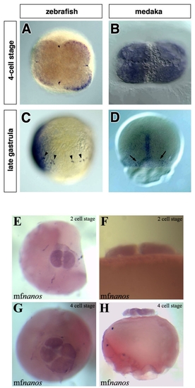
The early expression patterns of zebrafish-vasa, medaka-olvas and medaka-nanos1 mRNAs. In zebrafish, the vasa transcript is enriched at the marginal positions of the first two cleavage planes (A), while at a similar stage olvas mRNA is uniformly distributed throughout the cytoplasm of all blastomers (B). Throughout development in zebrafish, the vasa mRNA is expressed exclusively in the PGCs that can be found in random dorsoventral positions during blastula and gastrula stages (arrowheads in C). In contrast, at the first time point when medaka PGCs can be observed (stage 16), they are found on both sides of the embryonic shield on the dorsal side of the embryo (arrows in D). All images shown are whole-mount in situ hybridizations. The probes used are: vasa (A), vasa and chordin (C), olvas (B, D) and nanos1 (E to H). A, B, E and H are animal view, C is a lateral view with the dorsal aspect (labeled with chordin) to the left and D is a dorsal view, F and H are lateral views. |
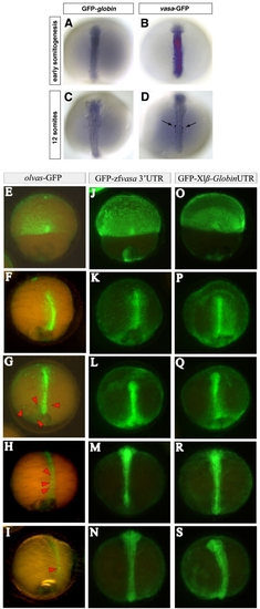
Stabilization of zebrafish vasa and translational regulation of medaka olvas mRNA in PGCs of medaka. 100 pg of GFP tagged fusion RNAs were injected at the 2-cell stage. (A to D) The embryos were fixed at early somitogenesis and at 12-somite stage and in situ hybridization using a GFP antisense RNA probe was performed to reveal the spatial distribution of the injected RNAs. In the control embryos the GFP-globin RNA is degraded uniformly in all cells (A and C) whereas in vasa-GFP injected embryos (B and D), the PGCs retain the injected RNA (arrows in D) while it is degrading in somatic cells. (E to S) olvas-GFP (E to I) as well as GFP-zfvasa 3′UTR (J to N) constructs were injected in medaka and followed for GFP expression and compared to control GFP-β globin 3′UTR injected embryos (O to S). Interestingly, in medaka, olvas-GFP protein expression was rapidly restricted to PGCs (E to I), while zebrafish vasa 3′UTR did not induced selective GFP expression. |
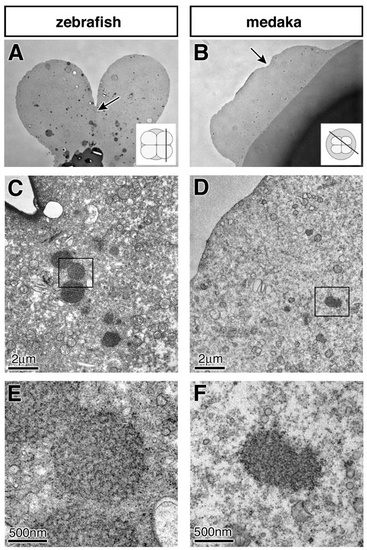
Sections of early 4-cell stage zebrafish and medaka embryos. Semi-thin section of zebrafish (A) and medaka (B) embryos visualized in a light microscope. Arrows indicate the areas shown in C-F representing the cleavage positions. The insets represent the orientation of the sections. Low magnification at the electron microscope showing the region of the germ plasm of zebrafish (C), and the corresponding position in medaka (D). At a higher magnification, the germ plasm of zebrafish is visible as distinct amorphous inclusions with a regular spherical shape and a filamentous fine structure lacking a surrounding membrane (E). At this magnification germ plasm resembling structure composed of irregularly shaped amorphous inclusions with a granular fine structure can be observed in medaka embryos. As described for zebrafish [28], the putative germ plasm of medaka can be followed in serial sections. |
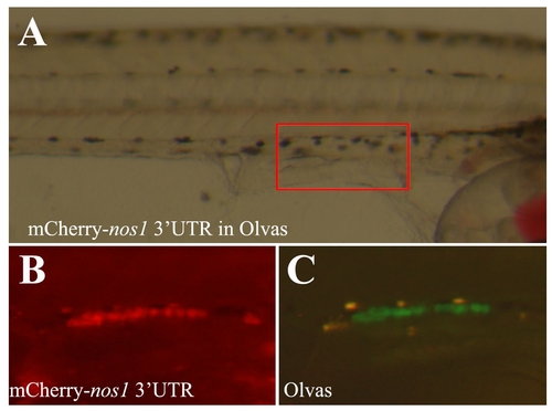
Zebrafish nanos1 3′UTR drives PGC-specific GFP/RFP expression. To confirm Zebrafish nanos1 3′UTR driven PGC-specific expression, the RFP- Zfnos1 3′UTR construct was injected in the Medaka Olvas transgenic strain (A) expressing GFP under the control of the vasa-promoter [45]. Overlap of GFP (B) and RFP (C) indeed confirm PGC specific expression under the control of Zebrafish nanos1 3′UTR. |