
Fig. 4
- ID
- ZDB-FIG-110407-4
- Publication
- Padanad et al., 2011 - Pax2/8 proteins coordinate sequential induction of otic and epibranchial placodes through differential regulation of foxi1, sox3 and fgf24
- Other Figures
- All Figure Page
- Back to All Figure Page
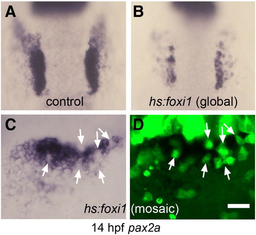
Misexpression of foxi1 inhibits otic development. (A, B) Expression of pax2a at 14 hpf in a control embryo (A) and a hs:foxi1 transgenic embryo (B) heat shocked at 11 hpf. (C, D) Expression of pax2a at 14 hpf in a mosaic embryo as seen under bright field (C) and fluorescence imaging (D). The mosaic was produced by transplanting lineage-labeled hs:foxi1 transgenic cells (green fluorescence) into a non-transgenic host. The embryo was heat shocked at 11 hpf to activate the transgene. Note the absence of pax2a expression in transgenic cells (white arrows). Images show dorsal views with anterior to the top (A–B); lateral views with anterior to the left (C–D). Scale bar, 50 μm (A, B), 25 μm (C, D). |
| Gene: | |
|---|---|
| Fish: | |
| Condition: | |
| Anatomical Term: | |
| Stage: | 10-13 somites |
| Fish: | |
|---|---|
| Condition: | |
| Observed In: | |
| Stage: | 10-13 somites |
Reprinted from Developmental Biology, 351(1), Padanad, M.S., and Riley, B.B., Pax2/8 proteins coordinate sequential induction of otic and epibranchial placodes through differential regulation of foxi1, sox3 and fgf24, 90-98, Copyright (2011) with permission from Elsevier. Full text @ Dev. Biol.