
Fig. 5
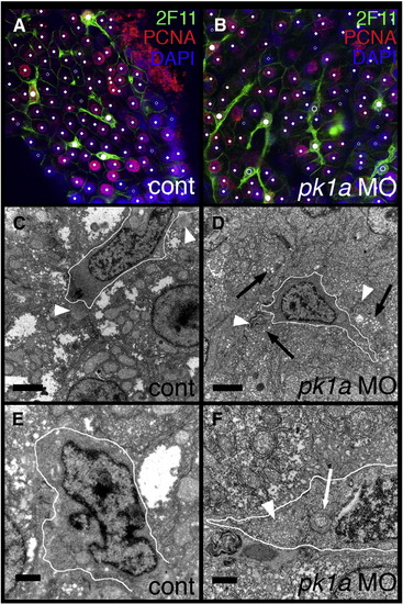
Abnormal bile duct cells in pk1a morphant livers. (A–B) Single confocal optical slices of whole-mount 2F11 and PCNA immunostaining of livers from 5 dpf control (A, cont) and pk1a morphant (B) larvae. 2F11 staining is in green, PCNA in red, and DAPI counterstain in blue. Hepatocytes are noted with small white dots, while biliary cells are noted with larger dots. Solid dots represent cells that are PCNA positive, while the open dots are PCNA negative. Note that the biliary cells in control are all PCNA positive, while there are PCNA negative biliary cells in the pk1a morphant. (C-F) Electron micrographs from livers from control (C, E, cont) and pk1a morpholino-injected (D, F, pk1a MO) larvae. (C–D) Low power views (scale bar 2 μm) demonstrate overall similarity in appearance, but canaliculi (white arrowheads) appear to have accumulated material within in the morphants (D), and there is an accumulation of vesicles (black arrows) in the hepatocytes in (D). (E–F) Higher power views (scale bar 500 nm) demonstrate dilated Golgi (white arrow) in the bile duct cell in the morphant sample (F), as well as an accumulation of intracellular vesicles (F, white arrowhead). The white outlines circumscribe the bile duct cells in C–F. |
| Fish: | |
|---|---|
| Knockdown Reagent: | |
| Observed In: | |
| Stage: | Day 5 |
Reprinted from Developmental Biology, 351(2), Cui, S., Capecci, L.M., and Matthews, R.P., Disruption of planar cell polarity activity leads to developmental biliary defects, 229-241, Copyright (2011) with permission from Elsevier. Full text @ Dev. Biol.