
Fig. s3
- ID
- ZDB-FIG-110322-29
- Publication
- Ogura et al., 2009 - Adaptation of GAL4 activators for GAL4 enhancer trapping in zebrafish
- Other Figures
- All Figure Page
- Back to All Figure Page
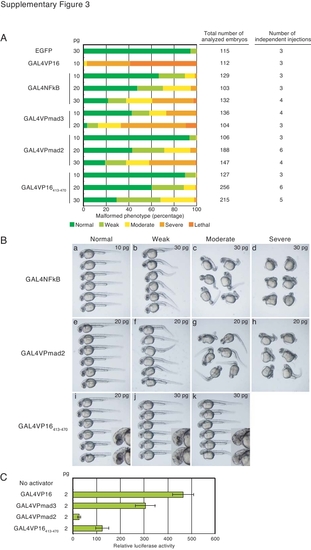
Supp. Fig. 3. Comparison of the toxicity and transactivation potentials of the variant GAL4 activators including GAL4VP16413-470 in zebrafish embryos. A: Comparison of the toxicity levels of the various GAL4 activators. The indicated GAL4 activator mRNAs (10, 20, or 30 pg) were injected into one- to two-cell-stage embryos (TL line) and morphological abnormalities at 28–30 hpf were classified according to their severity into five groups: normal, weakly malformed, moderately malformed, severely malformed, and lethal phenotypes. The affected tissues varied depending on the activators as shown in B. EGFP mRNA (30 pg) was also injected as a control. B: Typical morphologies for each phenotypic class. GAL4NFkB (a, 10 pg; b–d, 30 pg), GAL4VPmad2 (e–h, 20 pg), and GAL4VP16413-470 (i, 20 pg; j,k, 30 pg). Because GAL4VP16413-470 caused defects in the eye and the anterior CNS (insets of j, k) without significantly affecting posterior development, the anterior phenotype was mainly used as a criterion of phenotypic classes. C: Comparison of the transactivation potentials of GAL4VP16, GAL4VPmad3, GAL4VPmad2, and GAL4VP16413-470. Co-injection of GAL4 activator mRNAs (2 pg) with the UAS:Venusluc reporter vector was carried out as described in Figure 3. A low amount of activator mRNAs was used, as the adverse effects of GAL4VP16 are negligible at this expression level. |